Exploration and diagnostic techniques
We perform a wide range of tests and studies of high quality and precision


CONTRAST-ENHANCED MAMMOGRAPHY

Locate precisely
all suspicious injuries

Helps distinguish between
benign and malignant lesions

Reduces the
number of biopsies

We save time by
diagnosis of breast cancer
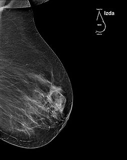
Contrast-Enhanced Mammography is the most accurate and comfortable diagnostic test for the detection of Breast Cancer. It is especially recommended for women with dense breasts, women who have already suffered the disease, and women with a family history of Breast Cancer. Contrast-Enhanced Mammography replaces standard Mammography and is performed in a similar way to it, with the addition that contrast is administered intravenously to the patient two minutes before the test.
The examination lasts approximately 3 minutes and is carried out with the patient standing. At IMAGINE we deliver the results 15 minutes after taking the test and they are also discussed personally with the Doctor. Patients choose Contrast-Enhanced Mammography because it accurately locates all suspicious lesions, helps distinguish between benign and malignant lesions, reduces the number of unnecessary biopsies and accelerates the diagnosis of possible Breast Cancer, allowing for faster and more effective treatment of the illness.


MAMMOGRAPHY | Medical Areas


3D MAMMOGRAPHY TOMOSYNTHESIS
3D Tomosynthesis Mammography is an advanced breast imaging technique that provides detailed three-dimensional images of breast tissue, essential for the early detection of breast cancer. At our center, we use a specialized 3D mammograph that, with minimal radiation, captures detailed images in seconds through gentle compression of the breast. These images, evaluated by expert radiologists, offer accurate diagnoses even before the appearance of symptoms.
At IMAGINE we deliver results 15 minutes after taking the test and provide a detailed diagnostic report and advice on next steps, ensuring that each patient is well informed and prepared for the road to recovery.


MAMOGRAPHY | Medical Areas


BREAST BIOPSY AND STEREOTAXY
Breast Biopsy is a procedure in which a small sample of breast tissue is removed for further examination, especially when an abnormality is identified in your previously performed mammogram or physical examination.
Breast Stereotaxy is a precise technique used to locate and guide the Biopsy needle towards the area of interest with high precision, thanks to the use of mammographic images. This method is non-invasive, rapid and highly effective in diagnosing specific conditions, allowing for early and personalized intervention.
BREAST BIOPSY AND STEREOTAXY | Medical Areas


CT SCAN
CT SCAN (Computerized Axial Tomography) is a radiological technique that allows us to see the inside of the organs, obtaining multiple images that are processed and integrated into a final one. It is the basis for the diagnosis of many pathologies such as fractures, blood clots, hemorrhages, heart problems or in the detection of lesions suspicious of cancer. Data collection is very extensive and anomalies that do not appear on a simple x-ray can be determined.
CT scanning is quick, painless, non-invasive and accurate. At Imagine Barcelona we have a state-of-the-art SIEMENS Somatom SCOPE CT SCAN device that has allowed us to offer our patients a lower dose of low radiation than any other CT SCAN.
40% less radiation
50% less noise
Highest quality images


CT SCAN | Medical Areas


DIGITAL RADIOLOGY
Digital Radiology is the most common radiodiagnosis method that exists. At Imagine, we are committed to providing diagnostic services of the highest quality and we have cutting-edge equipment that allows the performance of all types of projections, as well as contrast-enhanced tests, such as hysterosalpingography, intravenous urography or esophagogastric transit, among others.
Our X-ray machine minimizes exposure time by reducing radiation by 30% and offers high-quality images in just 2 seconds. This advancement not only improves diagnostic accuracy, but also enhances the patient experience by reducing the time needed to perform tests.


DIGITAL RADIOLOGY | Medical Areas


ORTHOPANTHOMOGRAPHY AND DENTAL CT
Orthopantomography is a radiographic technique that offers a panoramic view of the mandible, maxilla and teeth in a single image. It allows dentists to visualize the complete state of the patient's oral structure and helps detect problems such as hidden cavities, infections, root abnormalities and other oral problems, facilitating accurate diagnosis and timely treatment.
Dental CT is an advanced imaging technique that provides detailed, three-dimensional views of the teeth, jaw, nerves and surrounding soft tissue. This technology is particularly useful for evaluating complex problems, planning dental implants, identifying oral pathologies, and analyzing dental and bone anatomy in great detail.

ORTHOPANTHOMOGRAPHY AND DENTAL CT | Medical Areas


ULTRASOUND, DOPPLER AND FIBROSCAN
Ultrasound is an ultrasound diagnostic method that generates images of the structures inside us. It has multiple applications: diagnoses diseases of the gallbladder, prostate or genital problems, as a complementary test to mammography (breast), to evaluate metabolic bone diseases or inflammation in the joints, to visualize the uterus and ovaries during pregnancy, among others.
Combined with Color Doppler and Power-Doppler Ultrasound, we can see the flow of blood. In this way we can evaluate and carry out vascular studies of the lesions.


FIBROSCAN
The FibroScan is an essential non-invasive medical tool for evaluating liver health, identifying conditions such as liver fibrosis and cirrhosis quickly and accurately. Using ultrasound elastography, it measures the stiffness of liver tissue by calculating the speed of shear waves, offering a painless and efficient alternative to invasive biopsies.
In just a few minutes, patients obtain quantitative results that aid in the timely diagnosis and management of chronic liver diseases, ensuring a personalized care approach and minimizing the need for invasive procedures.
ULTRASOUND, DOPPLER AND FIBROSCAN | Medical Areas


BONE DENSITOMETRY
Bone Densiometry is a diagnostic test used to determine Bone Mineral Density (BMD). Our densitometers are low radiation and calculate the density of bones at different points of the skeleton. Thanks to this test we predict the risk of suffering fractures due to osteoporosis, as well as detect structural changes that become more fragile bones. We recommend the study to people with a family history of osteoporosis or who suffer from diseases related to bone loss, hypoparathyroidism and hyperparathyroidism.


